Training on Faster COVID-19 Testing
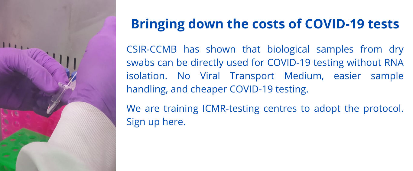
CSIR - Centre for Cellular & Molecular Biology
Council of Scientific and Industrial Research
Ministry of Science & Technology, Govt. of India
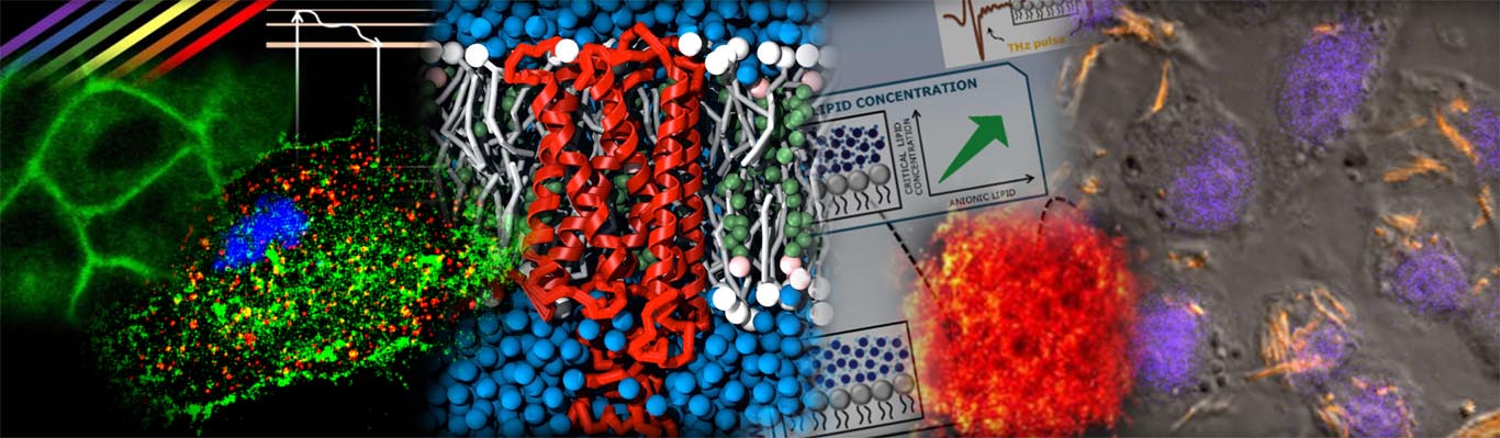
Integrative structural biology is a powerful approach for understanding the structural and functional properties of biomolecules, and for advancing the field of drug discovery. To obtain a more thorough knowledge of biological structures and processes, it integrates numerous methods such as X-ray crystallography, nuclear magnetic resonance spectroscopy, and cryo-electron microscopy. Furthermore, biophysical characterization of proteins using techniques such as isothermal titration calorimetry (ITC), surface plasmon resonance (SPR), differential scanning calorimetry (DSC), dynamic light scattering (DLS), and analytical ultracentrifugation (AUC) is critical for understanding their structures, interactions, stability, dynamics, and aggregation. In summary, these methods can be used to study the interactions between molecules, understand how they function in cells, and develop new drugs and therapies. Additionally, integrative approaches also provide information about the dynamics of biomolecules, which can further help in understanding the mechanisms of their functions.
State-of-the-art and cutting-edge facilities for comprehensive biophysical and biochemical characterization as well as 3D structure determination are available at CSIR-CCMB.
They consist of:
The X-ray facility of Integrative Structural Biology provides state-of-the-art resources to elucidate the three-dimensional structures of macromolecules and their complexes at the atomic level. It is equipped with powerful microfocus rotating anode generators:1) MicroMax™ 007 HF (Rigaku) Cu anode generator with a Mar345-dtb image plate detector and Oxford cryosystem; and 2) FR-E+ SuperBright (Rigaku) dual-wavelength Cu/Cr anode generators with an R-axis IV++ image plate detector and X-stream cryosystem. The FR-E+ system is the most intense home-lab source available today for macromolecular crystallography, with focusing optics that can deliver a flux comparable to second-generation synchrotron beamlines. Data collected from single crystal diffraction is processed using crystallographic computational software. Structure determination and molecular modeling studies are performed on Intel Quad-Core windows and Linux-based workstations using software installed on the CCMB High Performance Computing (HPC) server.
High Throughput (HT) Crystallization
A state-of-the-art HT-Crystallization facility automates the complete crystallization experiment. The three major operational components are (i) Alchemist (Rigaku) for liquid handling, (ii) Protein Crystallization Robotic systems: Mosquito (SPT Labtech) and Oryx 4 (Douglas Instruments) for crystallization drop setting, and (iii) vibration-free rumed incubators(4 °C and 20 °C) for incubation and storage of crystal plates for crystal growth.
Dynamic Light Scattering (DLS)
The facility provides a dedicated Spectro Size-300 from NabiTech to determine the sample purity and size distributions of particles in solutions in the nanometer to micrometer range using dynamic light scattering (DLS). Dynamic light scattering is a useful tool for diagnosing the polydispersity, stability, and aggregation state of macromolecules in solution prior to crystallization and SAXS experiments.
Small Angle X-ray Scattering SAXS
X-ray facility is also equipped with in-house Small Angle X-ray Scattering (SAXS) System to decipher the physical and structural features of macromolecules in solution. SAXS allows to probe size, shape, quaternary structure and complex formation of molecules without crystallization. It helps in understanding (i) structural parameters [radius of gyration (Rg), maximum Dimension (Dmax), partial-specific volume (Vp) etc], (ii) dynamics of molecules and (iii) generation of low-resolution shapes of macromolecules.
The SAXS facility houses a BioSAXS-2000 (Rigaku) with 2-D Kratky collimation, mounted on the existing left port of the MicroMax™ 007 HF (Rigaku) Cu anode X-ray generator. It is equipped with an OptiSAXS Confocal Max-Flux (CMF) for higher brilliance at the sample position and data collection times in the range of minutes. The configuration incorporates an Automatic Sample Changer for unattended overnight operation and an Automatic Analysis Pipeline based on the ATSAS package from EMBL Hamburg.
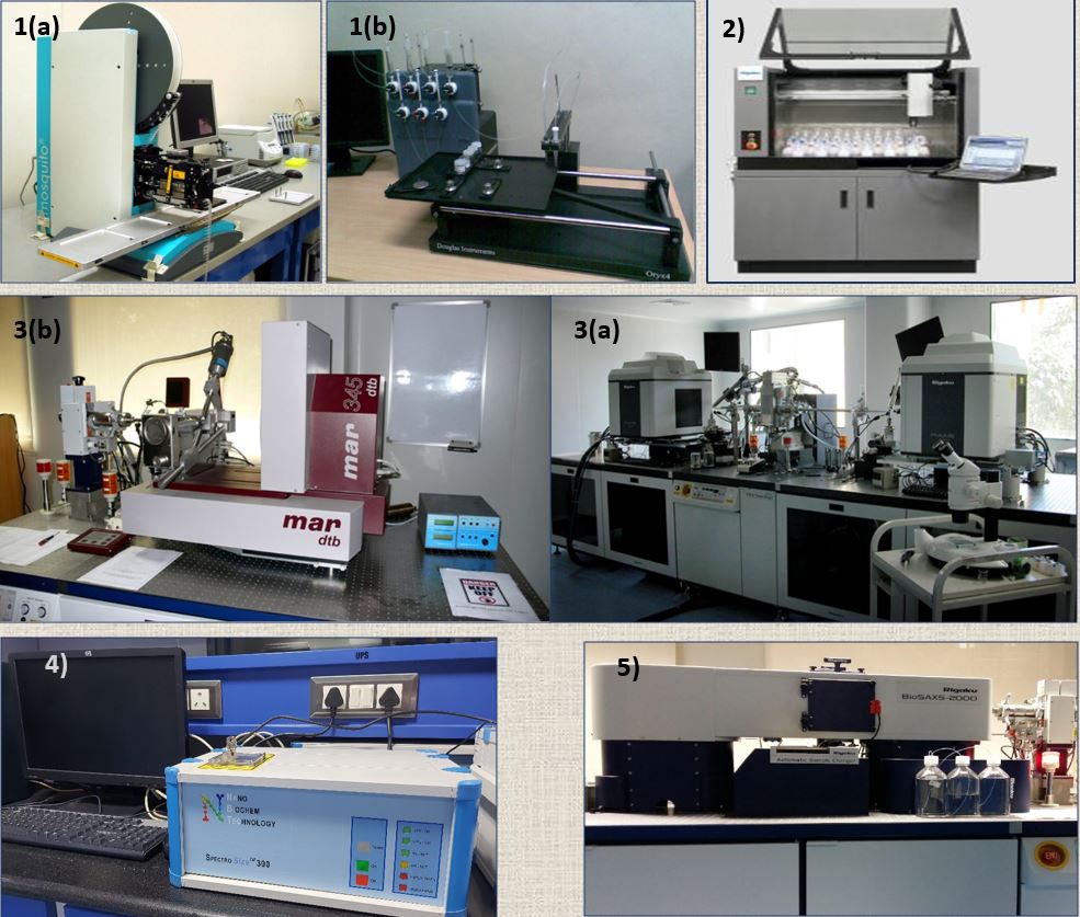
1) Robotic Systems: Automated crystallization: 1(a) Mosquito [TTP LabTech] Hanging Drop 1(b) Oryx 4 [Douglas Instruments] : Sitting Drop 2) Alchemist for liquid handling 3) X-ray diffractometers [Rigaku] 3(a) MicroMax™ 007 HF Rotating Cu anode generator with mar345 IP detector 3(b) FR-E+DW SuperBright System with Cu / Cr anode generators having R-axis IV++ image plate detector 4) Dynamic Light Scattering (DLS) [Nano Biochem Technology] 5) BioSAXS-2000[Rigaku] with 2-D Kratky collimation.
Several structural biology projects carried out at the CCMB and other research institutes or universities outside the CCMB and samples from industry/pharmaceutical companies are handled at these facilities.
For Use
Head-BDG
Phone: +91-40-27195541
Email: bdg@ccmb.res.in
Accessibility
Academic and Industries
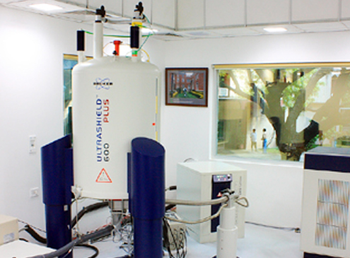
CCMB houses a state-of-the-art 600 MHz NMR Spectrometer equipped with a cryogenically cooled probe.
Key strengths
Detailed description
A 600 MHz narrow-bore NMR facility was set up in 2009 to study the biomolecular structure and function under physiological conditions in solution. The facility consists of a four channel NMR spectrometer equipped with a cryogenically cooled probe. The enhanced sensitivity of the cryoprobe allows de novo 3D structure determination of relatively large proteins (MW > 25 kDa) and nucleic acids as well as their ligand-bound complexes at the physiological condition. In addition, the spectrometer is routinely used to study dynamic events occurring in the biomolecules at a variety of timescales.
This facility is useful for performing structural studies of dynamic biomolecules that are difficult to crystallize (e.g., multi-domain proteins and majorly disordered proteins). The spectrometer is routinely used to derive biologically relevant conformational flexibility of proteins and nucleic acids in situ. Some of the important findings derived from the data generated by the facility are: (1) The solution structure of RDE-4 (C. elegans) elucidated structural modifications in both dsRBDs that were responsible for selecting the trigger dsRNA (2) Understanding the RNAi initiation in plants through the solution structure complemented with the structure-based activity assays of DRB4 (A. thaliana) (3) The solution structure of Crc (~32 kDa and presumably the largest solution structure derived by NMR from India) revealed its non-canonical RNA binding surface responsible for regulating the carbon catabolite repression process (4) Understanding the process of enantioselection to elucidate the mechanism of chiral proofreading during protein translation.
Over the years, NMR (Structural Biology) has become an integral part of CCMB’s research activities and has immensely contributed to numerous projects including studies and design of thermostable lipases, studies on antimicrobial peptides, study of the interaction of intracellular loops of GPCRs with membranes, and structure-function relationships of key proteins in P. falciparum, among others. The data generated by the NMR facility has been used in research articles published from CCMB in several internationally acclaimed scientific journals such as Proc. Natl. Acad. Sci. USA (2010), J. Mol. Biol. (2011), eLife (2013), Biochem. J. (2014), PLOS Biol. (2016), Nucl. Acids Res. (2017), and J. Magn. Res. Open (2022).
For Use
Head-BDG
Phone: +91-40-27195541
Email: bdg@ccmb.res.in
Accessibility
Academic and Industries
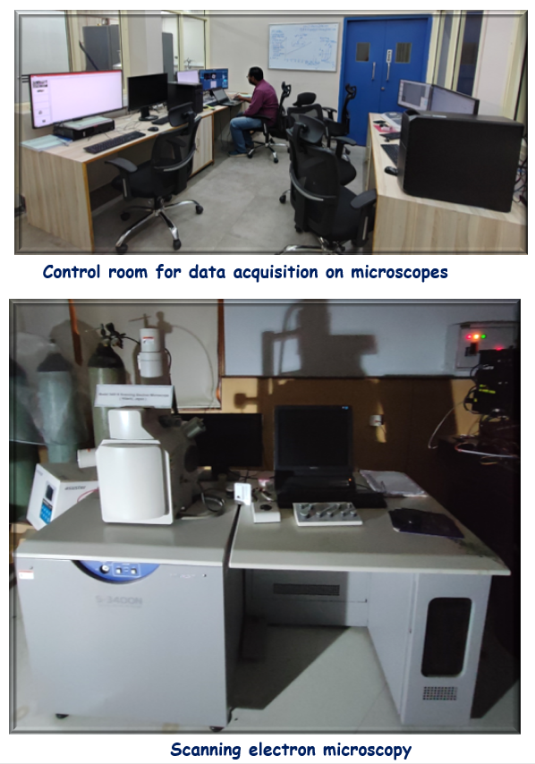
The electron microscopy centre at CSIR-CCMB was established in March of 2022 to help researchers understand the molecular underpinnings of life at unprecedented details. The centre is equipped with state-of-the art cryogenic transmission electron microscopes with advanced cameras capable of high throughput imaging of biological specimen at near native conditions and thin sections of biological specimens. The facility is also equipped with sophisticated high precision sample preparation equipment.
This centre will help researchers in delineating the underlying cause of diseases and development of new therapeutics and drugs for cure. This facility is not only being used by CSIR-CCMB scientists, but also by users from other institutes in India and pharmaceutical industries.
Transmission Electron Microscopes
Talos L120C Transmission Electron Microscope
Talos L120C is a 20-120 kV cryogenic transmission electron microscope with a LaB6 filament electron source and also equipped with a fast readout Ceta camera. This scope is used for imaging biological specimens at room temperature or cryogenic conditions.
Transmission electron microscope (200 kV)
The 200 kV Talos Arctica cryogenic electron microscope has an x-FEG electron source and is equipped with Falcon IV direct electron detector and CetaD cameras for ultra-high-resolution structure determination. This has a robotic autoloader system which is capable handling up to 12 vitrified grids. High-throughput, automated imaging is performed using the EPU software suite.
Manufacturer: Thermo Fisher Scientific
Model: Talos Arctica G2

Additional Instruments for Sample preparation in electron microscopy centre
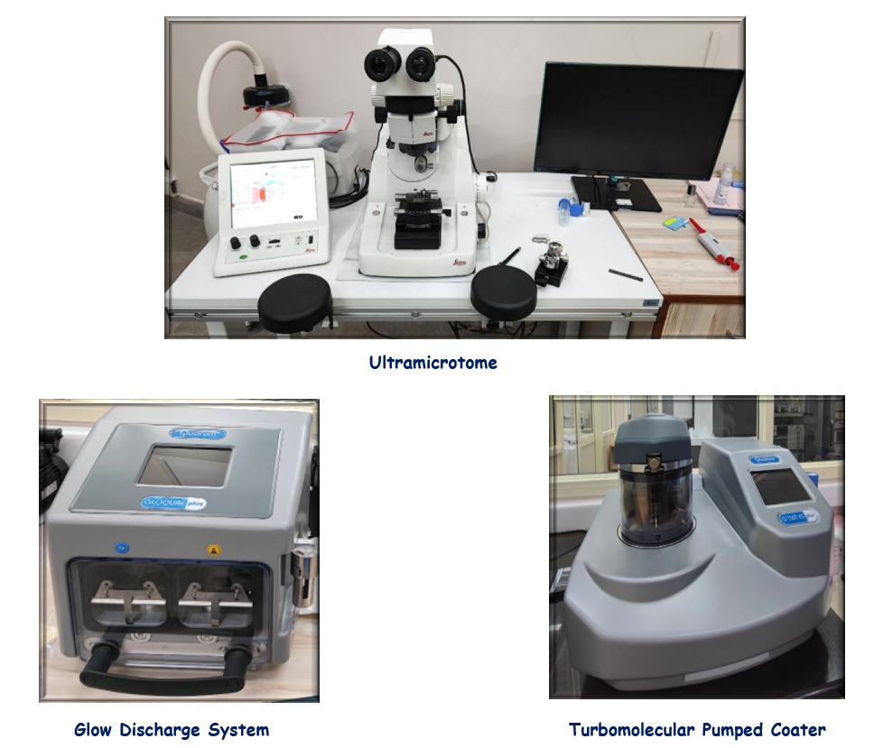
Ultramicrotome
To obtain high-quality, ultrathin (~100nm thickness) sections of biological specimen at room or cryogenic temperatures, the centre houses a low-temperature sectioning ultramicrotome system.
Manufacturer: Leica Microsystems
Model: Ultramicrotome Leica EM UC7 with FC7
Glow Discharge System
Glow discharge unit for cleaning, hydrophilization and surface modification of TEM Grids. This system is equipped with two chambers which helps researchers to choose discharge in-air or in-chemical vapor.
Turbomolecular Pumped Coater
This turbomolecular pumped coater operates at lower vacuum. This enables the sputtering of metals such as gold, platinum etc. with a lower grain size. This makes sputter coated samples suitable for high-resolution imaging.
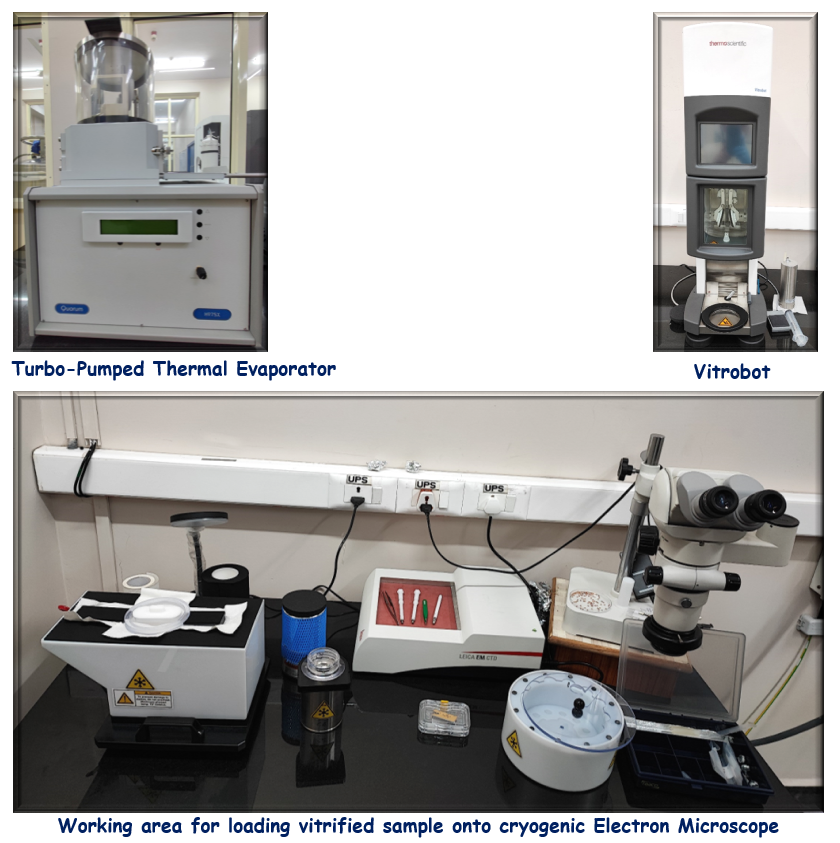
Turbo-Pumped Thermal Evaporator
A compact, bench-mounted, thermal evaporator for vacuum deposition of thin layers of carbon (for preparation of ultra thin carbon support films).
Vitrobot
The Vitrobot system is a robotic system used for preparation of thin vitrified grids under precise temperature and humidity controlled conditions for high-resolution cryogenic electron microscopy. Precise ice thickness of vitrified samples can be achieved by controlled blotting parameters in this robot and it not only improves sample quality, but also provides reproducibility of cryogenic sample preparation.
For Use
Head-BDG
Phone: +91-40-27195541
Email: bdg@ccmb.res.in
Accessibility
Academic and Industries
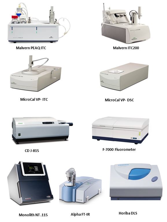
The facility offers a large panel of state-of-the-art equipment to address queries regarding the structures, folding/stability, and interactions of biomolecules. Proper hands-on training is provided to users, and equipment time may be booked around the clock by mail to the Business Development Group (BDG) or through the AnalytiCSIR portal (for academic and industrial users). The BCF is managed by a PhD scientist who assists researchers in the design, implementation, and analysis of experiments and results. In addition, the instrumentation division of the CSIR-CCMB supports the maintenance and repair of various instruments, which reduces the downtime of instruments.
The BCF is equipped with spectroscopic and calorimetric instruments for the study of structure, folding, binding thermodynamics and kinetics, and quantitation experiments. For biomolecular interactions, BCF offers multiple calorimetric instruments such as PEAQITC, ITC200, VP-ITC, and VP-DSC, and new instruments such as Nano Temper Monolith. N115
To determine the secondary structure, the facility features a Jasco J-815 circular dichroism spectrometer with Peltier-based temperature control, a Horiba dynamic light scattering (DLS) instrument for quick molecular size characterization, zeta potential, and Brucker’s Alpha FT-IR spectrometer for powdered samples. Various high-end fluorometers are available to measure the fluorescence lifetime as well as regular excitation/emission spectra. The complete list of instruments is as follows:
Spectroscopy/Fluorometry
Other Instruments
For Use
Head-BDG
Phone: +91-40-27195541
Email: bdg@ccmb.res.in
Accessibility
Academic and Industries

 CSIR-CCMB Aug-2025 PhD Program: List of selected candidates